
Many patients are familiar with 4-D ultrasound diagnostics in the field of gynecology. It is used here to represent the fetus in the womb in the best possible and most detailed way. This creates images like the following:
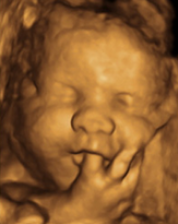
(Source: www.9Monate.de)
We use these procedures to display your vessels and organs in detail. With that, we have a precise idea of your overall
situation and are able to clearly visualize and explain this to you.
What exactly happens with ultrasound?
From the point of view of physics, ultrasound is understood to mean sound waves outside the sound frequencies perceivable for the human ear. During medical ultrasound examination, this form of
acoustic energy is sent into the body and reflected there by the organs, etc. These reflections are collected by the device again and then converted into a visualization. This conversion of sound
into images is called "sonography".
Today's ultrasound technology enables a detailed and, above all, risk-free and side effect-free representation of the
internal organs of humans and, due to its gentle approach, has played an important role in general diagnostics for many years. It comes to the first detection of degeneration in the organ or
vascular area, as well as the detection of pregnancy e.g. for use.
The 3D ultrasound
In contrast to the two-dimensional representation, a 3D ultrasound now also enables a spatial, three-dimensional representation of your organs and vessels. With a movable probe, normal ultrasound
reflections are put together from a specially developed computer to form a three-dimensional image.
Our 4D ultrasound
4D ultrasound is also understood to mean a three-dimensional ultrasound examination under real-time conditions. Instead of just displaying static images, a 4D ultrasound records the movements
within your body, which is why it is also called live 3D ultrasound. This approach enables extremely photo-realistic images, in which liquids and organ movements can also be captured in moving
sequences (4D).
The examination, like normal ultrasound, can be carried out almost at any time.
We would be happy to take a look at your "inside" together with you in the INUS MEDICAL CENTER.
